Na webu probíhá technická aktualizace systému a uživatelských dat. Některé funkce, včetně přihlášení, mohou být dočasně omezeny.Děkujeme za pochopení.
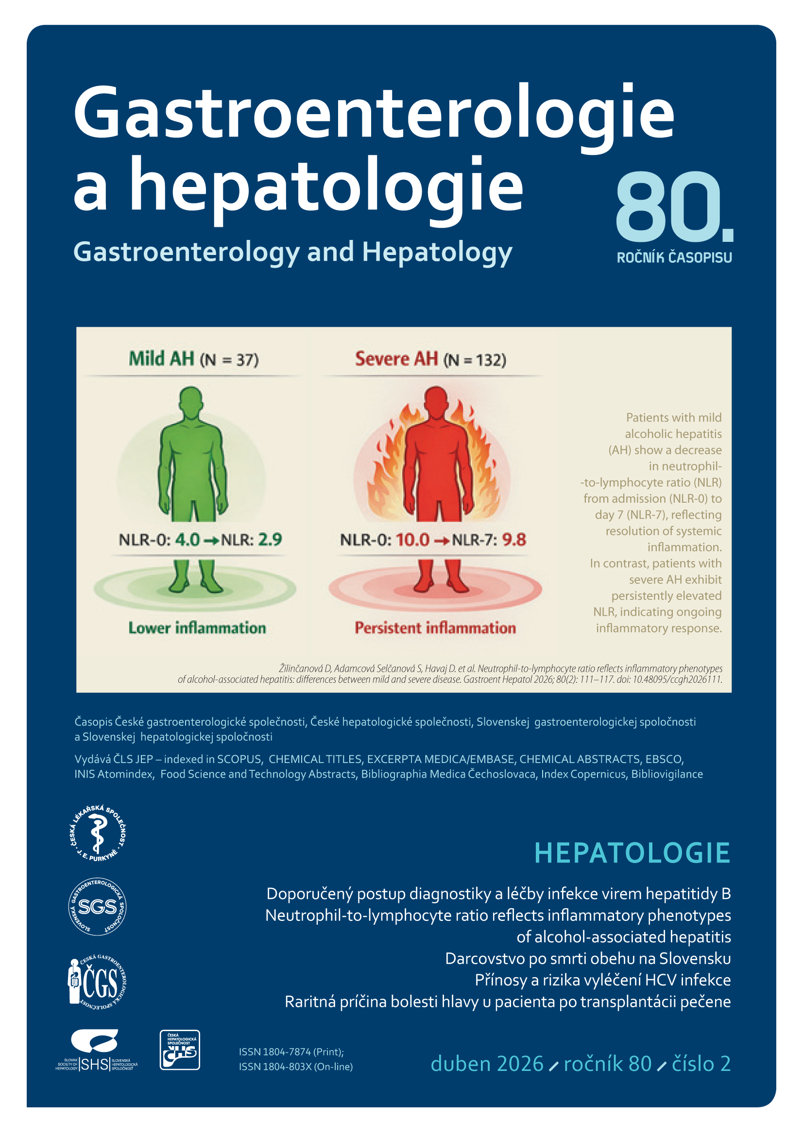
HEPATOLOGIE
Více…
Video - 1. díl roku 2026