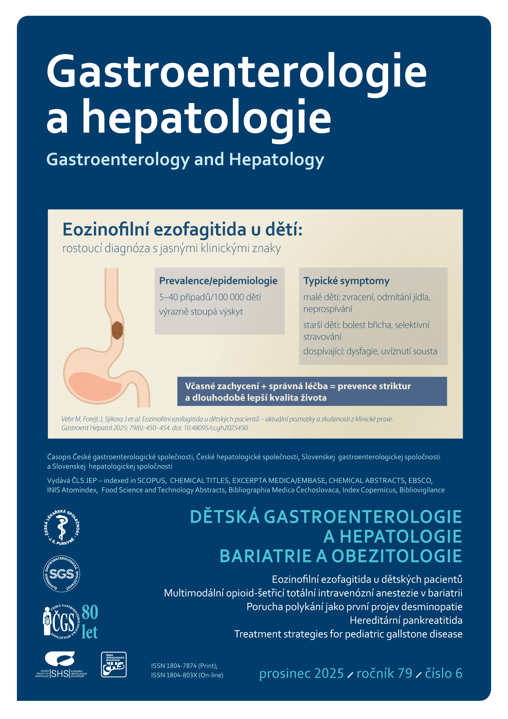
Anonymous User
Login / Registration

Gastroenterologie
a hepatologie

Gastroenterology and Hepatology

Gastroent Hepatol 2018; 72(6): 473–478. doi:10.14735/amgh2018473.

Evaluation of liver steatosis in obese paediatric patients using ultrasonography

Marek Pršo1, Marek Kozár2, Terézia Kráľová1, Zuzana Michnová1, Zuzana Havlíčeková Orcid.org  3, Lýdia Zúbriková1, Peter Bánovčin Orcid.org  1

+ Affiliation

Summary

In healthy children, tissue echogenicity of the liver parenchyma is comparable to that of the kidneys or spleen. With obesity, the most prominent risk factor for non-alcoholic fatty liver disease (NAFLD), overall liver tissue echogenicity increases with the severity of steatosis. Currently, ultrasonographic quantification of liver steatosis using the hepatorenal index (HRI) is coming to the forefront. The HRI value is defined as a histogram ratio between tissue echogenicities of the liver and those of the right kidney, and its increase is proportional to the severity of steatosis, while overall liver tissue elasticity tends to decrease in more advanced stages of steatosis. In this study, we evaluated the significance and possible use of noninvasive ultrasonography for the assessment of the presence and stages of liver steatosis in paediatric patients with exogenous obesity. The obese children cohort showed higher HRI values than the healthy patient group (HRI = 1.43 ± 0.19 vs.  1.12 ± 0.07; p < 0,0001), and lower tissue elasticity than the control group (liver fibrosis index (LFI) = 1.64 ± 0.43 vs. 1.02 ± 0.27). The ultrasonograms also revealed that patients with higher body mass indexes and waist circumferences had more steatosis and lower liver elasticity. Liver elasticity was lower in obese patients with increased serum LDL and triglyceride levels, and the decrease also tended to be proportional to the decrease in serum vitamin D concentration. Based on these results, we conclude that obesity is a significant risk factor for the onset and development of NAFLD, and that noninvasive ultrasonographic methods can be used to diagnose and monitor obese patients with a high risk  of NAFLD.

Keywords

childhood, elastography, non-alcoholic fatty liver disease, obesity, ultrasonography

To read this article in full, please register for free on this website.

Benefits for subscribers

Benefits for logged users

Literature

1. WHO. Obesity and overweight. 2016. [on-line]. Available from: http: //who.int/media-centre/factsheets/fs311/en/.
2. Bodzsar EB, Zsakai A. Recent trends in childhood obesity and overweight in the transition countries of Eastern and Central Europe. Ann Hum Biol 2014; 41 (3): 263–270. doi: 10.3109/03014460.2013.856473.
3. Fedelešová M, Kupčová V, Szántová M et al. Možnosti použitia neinvazívnych metód pri nealkoholovej tukovej chorobe pečene. Gastroent Hepatol 2017; 71 (4): 325–332. doi: 10.14735/amgh2017325.
4. Koula M, Hrůzová D, Harmáček D et al. Metody neinvazivního stanovení pokročilosti jaterní fibrózy. Gastroent Hepatol 2016; 70 (2): 118–124. doi: 10.14735/amgh2016118.
5. Webb M, Yeshua H, Zelber-Sagi S et al. Diagnostic value of a computerized hepatorenal index for sonographic quantification of liver steatosis. AJR Am J Roentgenol 2009; 192 (4): 909–914. doi: 10.2214/AJR.07.4016.
6. Garg A, Misra A. Hepatic steatosis, insulin resistance, and adipose tissue disorders. J Clin Endocrinol Metab 2002; 87 (7): 3019–3022. doi: 10.1210/jcem.87.7.8736.
7. Duarte MA, Silva GA. Hepatic steatosis in obese children and adolescents. J Pediatr (Rio J)  2011; 87 (2): 150–156. doi: 10.2223/JPED.2065.
8. Akcam M, Boyaci A, Pirgon O et al. Importance of the liver ultrasound scores in pubertal obese children with nonalcoholic fatty liver disease. Clin Imaging 2013; 37 (3): 504–508. doi: 10.1016/j.clinimag.2012.07.011.
9. Hamaguchi M, Kojima T, Itoh Y et al. The severity of ultrasonographic findings in nonalcoholic fatty liver disease reflects the metabolic syndrome and visceral fat accumulation. Am J Gastroenterol 2007; 102 (12): 2708–2715. doi: 10.1111/j.1572-0241.2007.01526.x.
10. Vajro P, Lenta S, Socha P et al. Diagnosis of nonalcoholic fatty liver disease in children and adolescents: position paper of the ESPGHAN Hepatology Committee. J Pediatr Gastroenterol Nutr 2012; 54 (5): 700–713. doi: 10.1097/MPG.0b013e318252a13f.
11. Manco M, Bedogni G, Marcellini M et al. Waist circumference correlates with liver fibrosis in children with non-alcoholic steatohepatitis. Gut 2008; 57 (9): 1283–1287. doi: 10.1136/gut.2007.142919.
12. Michnová Z, Szépeová R, Havlíčeková Z et al. Prevalencia hepatopatie u adolescentov s diabetes mellitus 1. typu. Pediatria 2017; 12 (Suppl 2): 53.
13. Saadeh S, Younossi ZM, Remer EM et al. The utility of radiological imaging in nonalcoholic fatty liver disease. Gastroenterology 2002; 123 (3): 745–750.
14. Oliveira AM, Oliveira N, Reis JC et al. Triglycerides and alanine aminotransferase as screening markers for suspected fatty liver disease in obese children and adolescents. Horm Res 2009; 71 (2): 83–88. doi: 10.1159/000183896.
15. Nobili V, Alisi A, Torre G et al. Hyaluronic acid predicts hepatic fibrosis in children with non-alcoholic fatty liver disease. Transl Res 2010; 156 (4): 229–234. doi: 10.1016/j.trsl.2010.05.008.
16. Cimini FA, Barchetta I, Carotti S et al. Relationship between adipose tissue dysfunction, vitamin D deficiency and the pathogenesis of non-alcoholic fatty liver disease. World J Gastroenterol 2017; 23 (19): 3407–3417. doi: 10.3748/wjg.v23.i19.3407.
17. Račanská E. Vitamín D – hormón, ktorý nám chýba. Prakt. lekárn. 2014; 4 (2–3): 53–55.
18. Havličeková Z, Szökeová A, Zúbriková L  et al. Aktuálny pohľad na nealkoholovú steatózu pečene v detskom veku. Pediatria 2015; 10 (3): 142–145.

Credited self-teaching test